Small Incision Cataract Surgery (Non Phaco SICS)
Abstract:
Purpose: To evaluate the technique and visual outcome of a prospective series of Non phaco Scleral tunnel sutureless Cataract surgery and I.O.L. implantation.
Methods: Nine hundred seventy cases of various types of cataract, which include 790 cases of senile cataract, 110 cases of complicated cataract, 50 cases of childhood cataract and 20 cases of traumatic cataract were taken for this study. Following sclerocorneal tunnel incision, capsulorhexis, hydro dissection, nucleus was delivered with the help of lens loop (microvectis). I/A was done with simcoe canula and lens implanted either in the bag or in the sulcus. In peadiatric patients, anterior chamber is reformed with air at the end of the surgery. Surgical complications, visual acuity at discharge, 2 weeks, 4 weeks and 6 months follow up are reported.
Results: Visual acuity corrected after 4 weeks was 6/18 or better in 95.97% of eyes in adult group, 54% in childhood cataract group. Complications like mild iritis is seen 80(8.24%) eyes, severe endothelial damage in 6(0.61%) eyes, intra operative complications like inferior iridodialysis noticed in 2(0.20%) eyes, damage to scleral spur and cyclodialysis in 3 eyes, hyphema in 4(0.41%) eyes, pc rent in 12(1.23%) eyes out of which vitreous loss in 8 eyes, IOL decentred in 3 eyes and IOL dislocated into vitreous in two eyes, which were removed intra-operatively. Posterior capsular opacification noticed in 82(8.45%) eyes, poor visual acuity noticed in 9 (1.03%) eyes in adult cataract group. One eye (0.10%) developed severe endophthalmitis.
Conclusion: SICS (Non-phaco) is our routine technique for all types of cataract, It involves nucleus expression by a microvectis of 4-5 mm size. The scleral tunnel incision varies from 6- 7.00 mm depending on types of cataract. Side port is necessary in difficult cases for rhexis and aspiration of sub incisional cortex. All the blades used are indigenous ones. Rapid visual recovery, and over all visual acuity corrected after 4 weeks is satisfactory. This suture less technique also extended to peadiatric cataracts(> 2 yrs of age), where A/C was reformed with air bubble at the end of the surgery. Considering it’s low cost and rapid visual recovery, we recommend this simple technique for all types of cataract in the developing countries.
Key words: SICS, Scleral tunnel, Non phaco, Manual phaco, IOL implantation.
Introduction:
Cataract surgery has witnessed a phenomenal progress over the years with addition of newer surgical technique and instrumentation. The methods of ICCE and ECCE, which we learned during our postgraduate training is perhaps no more in practice. Non phaco sutureless cataract surgery is the need of the hour in developing countries because, it is cost effective. Neither our patients nor the surgeons can afford phaco-emulsification (PE) procedure. Overall the technique of phacoemulsification (PE) is confined to a limited no.of surgeons in the developing countries because of the high cost of the equipment and it’s consumables. That is the reason why there is renewed interest in Non phacoemulsification small incision cataract surgery (Non Phaco SICS) in the third world countries. Several methods have been described for nucleus removal in SICS, in this procedure entire nucleus is removed through a sclerocorneal tunnel by a microvectis. We have been performing this technique in all types of cataract in our institute, Rajah Muthiah Medical College Hospital since last one decade, so far more than 10,000 small incision cataract surgeries have been performed including paediatric cases. This particular technique (microvectis) in SICS is evaluated and reported in this prospective study.
Materials and Methods:
Nine hundred and seventy consecutive cases of various cataracts, attending to ophthalmology OPC of R.M.Medical College Hospital, Annamalai University are the subjects of this study and all the cases operated by single surgeon (PM) were taken up for this study. This includes 790 cases of senile cataract, 50 cases of child- hood cataract and 110 cases of complicated cataract and 20 cases of traumatic cataract.Thorough preoperative evaluation of anterior segment of the eyes was done by biomicroscope, posterior segment by B scan ultrasonogram and indirect ophthalmoscope. The cases with coexisting posterior segment disorders were excluded from the study. The keratometry and I.O.L. power were calculated using Teknar Image 2000 A and B scan. The surgeries were performed under peribulbar anaesthesia except for childhood cataract, which were undertaken with general anaesthesia. Peribular anaesthesia was achieved by giving 2 injections, mixture of 5cc of 2% xylocaine with 5cc of 0.5% bupivacaine by two points technique without adding hylase. Facial N. block was avoided in all the cases. Ocular hypotony was achieved by ocular compression with pinky ball.
Surgical Method:
Scleral Tunnel Incision:
Following ab-externo fornix based conjunctival flap (Fig.1), a frown tunnel incision is given with B.P.blade no.15. We never use diamond knife for scleral incision and tunnel dissection, as it is very costly to maintain. The size of incision, the distance between the ends, mostly varies from 6.00 to 7.00mm, however if it is nuclear, rock hard cataract incision may be bigger up to 7.50mm. It is to be noted that the incision can be extended at any point of time. The anterior extent of the incision is always more than 2mm behind the limbus and two ends can be 4 to 5 mm behind the limbus. The tunnel was dissected (Fig.2) with the help of sharp edge tunnel (crescent) blade angled bevel up no 582620/582623.The incision depth is usually up to 2/3rd thickness of sclera. One should be extremely careful not to be very superficial or too deep to avoid complications like buttonhole or cyclodialysis. Further the thin flap has always the tendencies to tear. The internal incision, which is entry to anterior chamber, was made with the help of sharp edge sharp tip keratome, angled bevel upno.552620, 552820 and latter extended after capsulorhexis.
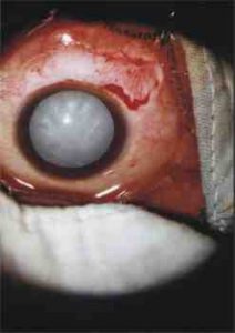
Fig. 1: Conjunctival flap reflected, hyper mature cataract

Fig.2: showing sclerocorneal tunnel dissection with crescent blade.
Capsulotomy:
Continuous curvilinear capsulorhexis, which was originally described, by both Gimbel (Canada) and Neuhann (Germany) has revolutionized the modern cataract surgery. This was performed with either 26G bent needle or with the help of Masket Capsulorrhexis forceps. Forceps is very useful in paediatric cases, because the capsule is very elastic in children. A rhexis of 6 to 7.0mm diameter is essential, how-ever if it is nuclear cataract, one or two relaxing incision are made at 2 0’clock and 10 0’clock meridians with the help of 26G bent needle. Whenever there is no red reflex, rhexis was easily performed by using trypan blue.
Hydro dissection:
This step is very essential before nucleus delivery. It is carried out with 2 ml syringe using curve 24G west lacrimal canula; the fluid was injected beneath the anterior capsule in one or two places, however large volume is avoided. Fluid wave or Golden ring reflex are observed to ensure complete hydro dissection and hydro delineation.
Nucleus management:
There are different techniques available for nucleus management in SICS, namely hydro-expression, phaco-sandwich, phaco-fracture, irrigating vectis etc. We restrict the discussion to microvectis technique only, as this is our preferred method of nucleus delivery in all types of cataract.
After reforming the anterior chamber with viscoelastic the superior pole of the nucleus was engaged, lifted up and rotated with the help of an I.O.L. dialer or 26G needle and subsequently prolapsed into the anterior chamber. Once the superior pole lifts up, viscoelastic may be injected underneath to make nucleus rotation easy. The nucleus rotation is done either clockwise, anti clockwise or both to luxate the nucleus completely into anterior chamber. Sometimes it is difficult to luxate the nucleus into anterior chamber in immature cataract. It is due to too much hypotony or incomplete hydrodissection , one has to reform the a/c with viscoelastic and do the hydrodelineation to overcome this difficulty. Similarly this problem may be encountered in nuclear cataract when the rhexis is small, where relaxing incision is given over the anterior capsule. Once the nucleus in anterior chamber, viscoelastic is placed both anterior and posterior to the nucleus. This step is essential to avoid endothelial damage. A microvectis or lens loop, 3-4 mm in size is introduced underneath the nucleus and the nucleus was expressed by gently applying forward pressure. This step is done in a more controlled fashion under direct visualization to avoid trauma to cornea and iris. Sometimes the epinucleus or portion of the cortex will be sheared off by the anterior lip of the incision. The remaining portion of cortex and epinucleus can be easily rotated and extracted by either by viscoexpression or aspirated by simcoe I/A canula. Visco-expression is carried out by injecting viscoelastic into a/c while depressing the posterior scleral lip. For easier removal of nucleus in nuclear cataract we recommend incision of 7-7.5mm to avoid intra operative complications, as stated earlier, some times two relaxing incisions over the anterior capsule, required in rock hard cataract to avoid complication like capsular tear or zonular dialysis. We never use anterior chamber maintainer nor irrigating vectis in this technique as we believe that viscoelastic protects the endothelium in a better way than BSS.
I.O.L. Implantation:
Viscoelastic material was injected into the capsular bag and in the anterior chamber before I.O.L. implantation. The I.O.Ls used were all indigenous, mainly Eye O’ Care, Appalens, and IMD and Cee On lenses with 5.5 to 6.5mm optics and 12.5mm to 13.5 mm overall diameter. In 370 eyes I.O.L.s were implanted in the sulcus and in 600 eyes, implanted in the bag. Viscoelastic was aspirated and anterior chamber was formed either with balanced salt solution, air bubble, or both, injected through the side port(Fig.3).
 |
 |
| Fig.3,4 Trypan blue assisted rhexis. | |
Closing the conjunctiva flap:
The conjunctival flap was closed with bipolar wet field cautery following subconjunctival injection. The sub conjuctival injection constitutes dexamethasone 0.5ml and cephazoline100mg, injected into the upper bulbar conjunctive, and eye patching applied with a pad. All these above mentioned procedures were carried out with the help of a zoom operating microscope.
Follow up
The patients were examined on the second day, 2nd week, 4th week and 6 months after the surgery. Slit lamp examination visual acuity, keratometry and indirect ophthalmoscopy were performed, any complications noticed were recorded. Reminder letters were sent to those who did not attend the regular follow up.
Results:
The mean age group of the patients was 57.8 yrs, the youngest child operated was 2yrs old. The no. of male patients was 378(38.96%) and no. of female patients was 592(61.04%). In adult cataract group nuclear cataract noticed in 24% eyes where as cortical type seen in 76% of eyes. In 96% of eyes the visual acuity corrected after one month was 6/18 or better in adult cataract group where as it is 54% in paediatric group. In 40(4.12%) eyes minimal corneal oedem was noticed, which subsided within 2 weeks of surgery. In 6(0.61%)eyes the best-corrected visual acuity was less than or equal to 6/60 because of severe corneal oedema. Three eyes had decentred I.O.L. and in other two implant dislocated in to vitreous cavity, which were explanted immediately. In one eye retinal detachment was noticed in the post operative period, which was pre-existing. Mild iritis was noticed in 80(8.24%) eyes and posterior capsular opacification was seen in 82(8.45%) eyes. Intraoperatively, posterior capsular rent was noticed in 12(1.23%) eyes out of which 8 eyes had vitreous loss, which was managed by anterior vitrectomy. In two eyes irido dialysis in the inferoior part with severe hyphema was encountered. In two cases of traumatic cataract the lenses were subluxated, which were removed through sclerocorneal tunnel, a/c IOL implanted in one and in other case scleral fixation IOL done following vitrectomy, visual acuity improved to 6/18 and 6/12 respectively. One eye (0.10%) developed severe endophthalmitis, we could not save that eye inspite of intravitreal antibiotics and vitrectomy.
Discussion:
The postoperative visual acuity, best corrected was 6/12-6/18 or better in almost 96% of cases at 4 weeks follow up in senile cataract group where as in paediatric group it is only 54%. In more than 80% of eyes in the adult group were having severely impaired vision before surgery, this was reduced to 1.03% at 4 weeks of surgery. In paediatric group, the percentage of good post operative visual acuity is low because there are so many factors, like age of onset of cataract, time of surgery, type of surgery and post operative management that affect visual prognosis in children. In 6(0.61%) eyes there were irreversible corneal oedema because of endothelial trauma. This complication will be much lower as one’s learning curve is overcome. The severe corneal oedema was due to endothelial decompensation, which was mostly seen in learning phase. In 2 (0.20%) eyes there were inferior iridodialysis and total hyphaema, which cleared slowly, and visual acuity improved to 6/24 and 6/36 after 2 months of surgery. The iridodialysis in the lower part was because of iris trapped in between microvectis and nucleus during delivery of nucleus. This can be avoided by injecting adequate amount of viscoelastic into the anterior chamber both above and below the nucleus so that free floating nucleus can be removed easily. In another two eyes there was zonular dialysis, implants were dislocating into the vitreous, which were explanted intra-operatively, vitrectomy was done and anterior chamber lenses were implanted. In other 12 (1.23%) eyes there were posterior capsular rent, out of which 8 had vitreous loss, which were managed by vitrectomy, IOL implanted in the sulcus in three eyes and in other five, it was implanted in the anterior chamber. The entire surgical procedure requires a good amount of surgical skill and additional training. Once the technique is mastered the surgery is easier, faster and the complication rate is low. In 4 eyes of complicated cataract there was posterior capsular plaques, it was difficult to aspirate, however in two cases re operation, I/A was done easily in the second post operative week, following which visual acuity improved to 6/12 and 6/18 respectively.
In early eighties Kansas described the phaco-fracture and Luther Fry, the phaco-sandwich technique for nucleus removal in SICS. Both these procedures are difficult because two instruments are introduced on either side of nucleus and there is every likelihood of endothelial damage either by instruments or by lens fragments. That’s the reason why we do not recommend any instrument in between nucleus and endothelium during nucleus delivery. Nucleus extraction with an irrigating vectis was first described by Steinert. Michael Bluementhal described nucleus delivery by using anterior chamber maintainer, he used to do all maneuvers underfluid, in both these procedure there is every chance of endothelial damage, because fluid never protects the endothelium in a better way. Richard Gianetti in 1996 reaffirmed that the nucleus capture is an inexpensive, phacoless, repeatable and relatively easy method of performing tunnel incision cataract surgery. He also stressed that no side port incision is required, surgeons can obtain the benefits of small self-sealing incision without the added cost of phaco. In first 200 cases of this study, no side port entry was done, however, at present we routinely do side port entry for easy capsulorhexis and aspiration of sub incisional cortex. We have observed that the endothelial loss is around 11%( our own unbublishrd data) in SICS, this usually occurs either during nucleus expression or during I/A. Lens fragments may touch the corneal endothelium during irrigation and aspiration.
Astigmatism is not so high as expected in spite of large incision in hard cataract, it has been observed that it is never more than 1.5-2D at 4 weeks follow up. The mean surgical induced astigmatism was 2.12 – 0.72 on the 2nd post operative week and reduced to 1.42 – 0.68 at the end of 4th week. In 87% of eyes the induced astigmatism was less than 1.50D. In our series most of the patient had against the rule astigmatism and showed faster astigmatic decay over time. We have observed that, even for incision of 8mm in nuclear cataract, there is no need of sutures, provided the incision is placed more posterior to limbus and tunnel is dissected longer, this also minimizes postoperative astigmatism. Longer(wider) the tunnel lesser the astigmatism it produces. Despite longer incision in some cases, it is a fact that we could achieve watertight, self-sealing, suture-less wound in almost 100% of cases of adult cataract and in children of more than 2 yrs of age. We routinely reform the anterior chamber with air bubble and BSS at end of the surgery in children to make it suture less.
Conclusion:
Non phaco small incision cataract surgery is useful in all types of cataract. It has contributed considerably to accelerated wound healing and minimisation of hospitalisation. The technique we advocate here is neither phaco-fracture nor phaco-sandwich, the nucleus is delivered by means of a microvectis, which is very simple procedure as it does not require bimanual technique. This tunnel cataract surgery is inexpensive, phacoless, relatively easy, repeatable and can be performed for any type of cataract from childhood to rock hard nuclear cataract. The wound is more secure with reduced intra-operative complications and virtually no chance of expulsive haemorrhage. Childhood cataract is much easier as there is no nucleus and in no time chamber collapses during surgery. One could achieve faster and satisfactory visual rehabilitation without added cost of phaco.This is indeed a safe and effective technique of sutureless cataract surgery in the hands of experienced surgeons. Considering the low cost and rapid visual recovery, we recommend this simple technique for all types of cataract in the developing countries.

Fig.5: A/C formed with air bubble following lens implantation, note: modified pocket tunnel.
References
- Girard L J, Hoffman RF. Scleral tunnel to prevent induced astigmatism. Am J Ophthalmol 1984; 97: 450-456.
- Kratz RP, Colveard DM, Mazzoceo TR,Davidson B. Clinical evaluation of terry surgical keratometer. Am Intraocular Implant Soc J 1990; 6: 249-251.
- Jack A Singer. Frown incision for minimizing induced astigmatism after small incision cataract surgery with rigid optic intraocular lens implantation. J Cataract Refract Surg 1991;17: 677-688.
- Mishra P. Small incision cataract surgery- Non Phaco SICS. Indian Intaocul Implant Refract J 2003;1:9-15.
- Kapoor Sashi. Incisions . Emmetropia, J Intraocular Implant and Refract Society 1999; 2: 17-25.
- Mishra P. Cataract surgery in children. Cyber lectures, www.indmedica.com/ophthal 2000; 1-5.
- Mody Kirit , Singh Gagan J. Small incision non-phaco cataract surgery. Emmetropia, J Intraocular Implant and Refract Society 1999;2:9-11.
- Kumar Ravindra. Small incision cataract surgery without phaco- my experience. Emmetropia, J Intraocular and Refract Society 1999; 2: 53-55.
- Mishra P. Microvectis Technique. In: Singh Kamaljeet,ed, Small Incision Cataract Surgery.Jaypee Brothers, NewDelhi, 2002,pp 113-116.
- Mishra P.Incision in Non phaco SICS, Orissa journal ophthal 2000; I I:30-32.
- Manual small incision cataract surgery, Aravind eye hospital, Madurai,2000.
- Luther F. The phacosandwich Technique. In Rozakis cataract surgery: Alternative small incision technique.Thorofare,N.J. Slack Inc.71-110.
- Kansas P. Phaco fracture In Rozakis cataract surgery: Alternative small incision technique. Thorofare, N.J. Slack Inc. 45-70.
- Hennig A, Kumar J,Yorston D et al. Sutureless cataract surgery with nucleus extraction. Br J Ophthalmol 2003;87:266-270.
- Thomas R, Kuriakose T, George R. Towards achieving small incision cataract surgery 99.8% of time. Indian J Ophthalmol 2000;48:145-51.
- Natchiar G. Manual small incision cataract surgery. Madurai, India: Aravind Publication,2000.
- Goel R, Malik KPS. Techniques of Nuclear delivery in Nonphaco small incision cataract surgery. Ophthalmol Today 2003;IV:92-94.
- Dandona L,Dandona R,Naduvilath TJ. Population based assessment of the outcome of cataract surgery in an urban population in southern India. Am J Ophthalmol 1999;127:650-658.
- Prajna NV, Chandrakanth KS, Kim R. et al. The Madurai intraocular lens study II: clinical outcomes. Am J Ophthalmol 1998;125:14-25.
- Holladat JT,Cravy TV, Koch DD, Calculating the surgically induced refracting change following ocular surgery. J Cataract Refract Surg 1992;18:429-443.
- Wright M, Chawla H, Adams A. Results of small incision extracapsular cataract surgery using the anterior chamber maintainer without viscoelastics. Br J Ophthalmol 1999;83:71-75.
- Peng Q, Hennig A, Vasavada AR et al. Posterior capsular plaque: a common feature of cataract surgery in the developing world. Am J Ophthalmol 1998;125-621-626.
- Haberle H, Anders N, Drosch S et al. Modification of no stitch technique in extracapsular cataract extraction by a single radial suture. Effect on post operative astigmatism.
- Morlet N, Minassian D, Dart J. Asigmatism and the analysis of its surgical correction. Br J Ophthalmol 2001;85:1127-38.
Correspondence:
Dr.P.Mishra, M.S. (Ophth),
Fellow Retina Foundation
Professor of Ophthalmology
RMMCH
Annamalai University-608002
Tamilnadu
e mail: [email protected]